2026-05-25
Rhinoplasty Revision - Case of an Excessively Long Implant (1)
A case study of a 20-year-old female undergoing rhinoplasty revision to correct a pinched nostril and drooping tip caused by an excessively long implant.
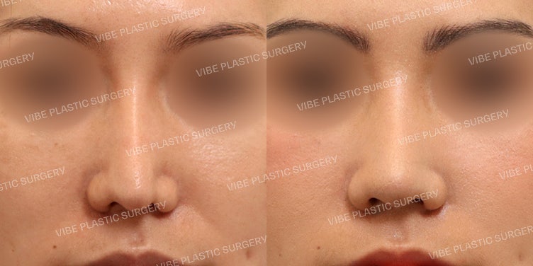
* Patient Information
# Female in her 20s
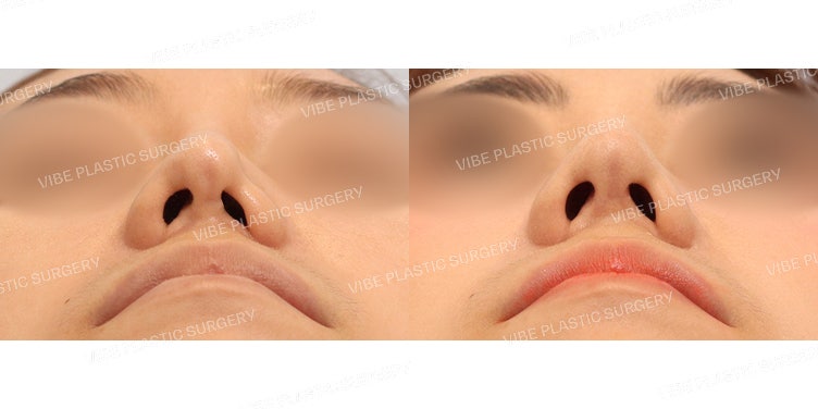
# Patient is concerned about severe deformation, as if one nostril is pinched, following a previous rhinoplasty.
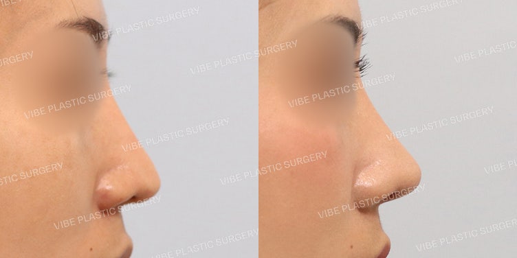
# The nasal tip has drooped and the columella has retracted, making the nose appear excessively long.
* Consultation
# A pre-operative CT scan confirmed that the implant was inserted too far down toward the nasal tip. It appears the implant was pressing against the tip, causing the deformation.
# Although the patient had tip surgery using septal cartilage previously, the support was weak, causing the columella to collapse and the tip to droop.
# The plan was to use donated rib cartilage to sufficiently elevate the nasal tip while correcting the deformed nostril and columella.
* Surgical Plan
# Nasal Tip Surgery (Correction of pinched nostril, long nose, and nasolabial angle)
# Bridge Surgery (Correction of deviated nose)
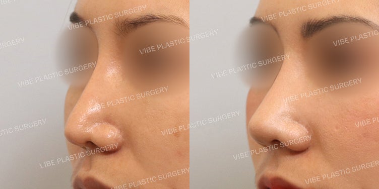
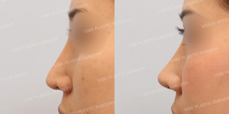
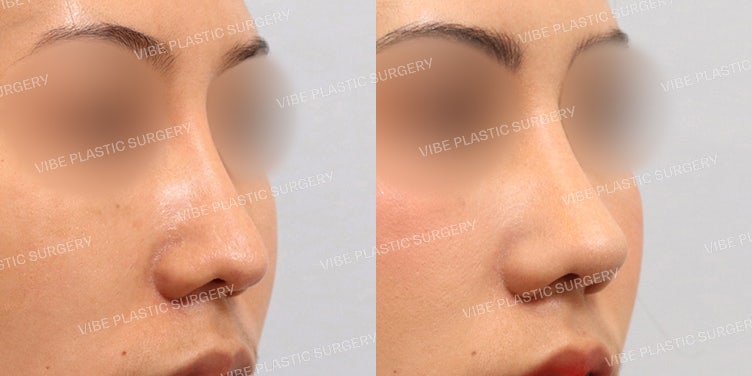
* Intraoperative Findings
# The implant extended too far into the tip, thinning the skin. Severe adhesion of scar tissue in this area caused the nostril deformation. The scar tissue was thoroughly removed, and the area was irrigated with an anti-scarring agent.
# The remaining septal pillar was too thin and lacked strength. A sturdy tip support was created using donated rib cartilage. The nasolabial angle was also corrected during this process.
* Post-operative Findings
# By correcting the tip deformation caused by the excessively long implant, an ideal nasal tip shape was achieved.
# When performing revision surgery, it is crucial to establish a perfect plan and prepare thoroughly beforehand. One must consider various variables in advance to respond flexibly to unexpected situations during surgery!
Always consult a board-certified plastic surgeon for consultations and procedures!
Wishing you health and beauty :)
Right Mind, Right Plastic Surgery - Dr. Yu Young-moon
Complications and side effects such as bleeding, inflammation, infection, and asymmetry may occur after surgery depending on the individual, and caution is required.
Frequently Asked Questions
What problems can arise if the implant is inserted too long?
Yes, if the implant is inserted too long, extending to the tip of the nose, it can press on the nasal tip and cause deformation. This can lead to the nasal tip dropping, the columella receding, making the nose appear longer, or in severe cases, one nostril may appear pinched. Additionally, the skin at the nasal tip can thin, and scar tissue may form.
What cartilage is used for revision rhinoplasty?
For revision rhinoplasty, various cartilages can be used depending on the patient's condition. In this case, because the remaining septal pillar from the previous surgery was thin and weak, donor costal cartilage was used to create a strong nasal tip support.
Can pinched nostrils be corrected?
Yes, pinched nostrils can be corrected through revision rhinoplasty. In this case, nostril deformation was observed due to thinning of the nasal tip skin and severe adhesion of scar tissue caused by the implant, and it was successfully corrected by removing the scar tissue and performing nasal tip plasty.
What examinations are performed before revision rhinoplasty?
Before revision rhinoplasty, a CT scan may be performed for accurate diagnosis. In this case, a pre-operative CT scan confirmed that the implant was inserted too long to the nasal tip and assessed the condition of the nasal septum, which helped in planning the surgery.
Can nasolabial angle correction be done simultaneously with revision rhinoplasty?
Yes, nasolabial angle correction can be performed simultaneously with revision rhinoplasty if necessary. In this case, the columella had collapsed during the nasal tip plasty, making the nasal tip appear dropped, and nasolabial angle correction was performed along with the construction of a nasal tip support using donor costal cartilage.